Intelligence That Sees More: How We Built an AI Model for Automatic Intrapulmonary Lymph Node Classification
Why This Problem Matters
Chest CT has become one of the most common diagnostic procedures worldwide. It identifies millions of pulmonary nodules — but only a minority represent cancer.
A significant portion of small solid nodules, up to 45%, are actually intrapulmonary lymph nodes (ILNs), also referred to as perifissural nodules (PFNs). These are normal anatomic structures that:
- often appear suspicious,
- may grow over time,
- cause concern for clinicians and patients,
- lead to unnecessary follow-up imaging and invasive procedures.
Yet numerous studies show: ILNs are not associated with lung cancer risk — even in oncologic populations.
However, their appearance varies widely, and radiologist assessment is highly subjective. Inter-observer agreement is only moderate, creating a need for a robust tool that can automatically and consistently classify these nodules.
What the Clinical Community Needed
Our mission was to create a solution that:
- reliably distinguishes ILNs from potentially malignant nodules,
- works both in screening and oncologic settings,
- reduces false positives,
- accelerates CT interpretation,
- brings confidence where uncertainty used to exist.
This is how the initiative to develop an AI model for ILN classification began.
How the Hiveomics Model Was Built
- Large Expert-Annotated Dataset
The model was trained on multiple datasets:
- internal Hiveomics clinical corpora,
- specialized research datasets,
- screening cohorts,
- oncologic patient datasets.
Annotations were performed by radiologists experienced in PFN morphology — crucial given the known variability in human interpretation.
- Formalizing PFN Morphological Criteria
We incorporated well-established CT criteria, including:
- proximity to pleura (≤10–15 mm),
- attachment to a fissure or thin septum,
- smooth, well-defined margins,
- oval, lentiform, or triangular shape,
- diameter up to 12 mm,
- absence of vascular connection,
- preference for lower-lobe locations.
These principles formed the structural backbone of the model.
- The Model Architecture
The Hiveomics AI combines:
- a 3D convolutional neural network optimized for small solid structures,
- a shape-analysis module,
- radiomic descriptors of local anatomy,
- a spatial-attention block highlighting pleural and fissural regions.
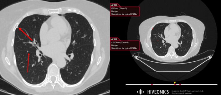
The Result: A Reliable Tool for Real-World Radiology
High Accuracy
The model achieves:
- AUC ~0.95,
- sensitivity >90%,
- specificity >85%.
These metrics match or exceed those reported in major international PFN-classification studies.
Reduced Interpretation Variability
Where radiologists often disagree, the model provides consistent results — reducing diagnostic uncertainty and improving decision-making.
Confidence in Difficult Cases
Even when a nodule:
- grows,
- lies against the pleura,
- appears in a cancer patient —
the model supports differentiation between benign and malignant lesions.
Seamless Integration Into the Hiveomics Ecosystem
The ILN classifier functions within the broader platform:
- full nodule detection,
- malignancy risk scoring,
- structured reporting.
Turning CT analysis into a structured and optimized process.
Conclusion
Developing an AI model for intrapulmonary lymph node classification marks a significant step forward:
- less uncertainty,
- fewer false positives,
- more efficiency,
- more confidence.
This is how we envision the future — where artificial intelligence empowers clinicians to see more than ever before.